


Internal Medicine
 The Internal Medicine Service at the Cornell University Hospital for Animals diagnoses and provides treatment for dogs and cats with a wide spectrum of health conditions. Our staff includes six board-certified veterinarians who have pioneered many of the approaches used for diagnoses and treatment of diverse canine and feline medical disorders used in veterinary clinics across the country. Internal Medicine faculty specialize in a wide range of health conditions including metabolic, infectious, endocrine, hematologic, gastrointestinal, liver, renal and respiratory disorders. Your pet will be seen by a veterinary team including faculty clinicians, attending residents, and students in training.
The Internal Medicine Service offers advanced diagnostic techniques including a wide range of minimally-invasive procedures. A collaborative working environment brings together the talent of a diverse group of veterinary specialty services including Anesthesiology, Oncology, Soft Tissue Surgery, Cardiology, Neurology, Imaging, and Clinical Nutrition to provide comprehensive veterinary care.
Advanced Techniques
- Arthrocentesis
- Balloon dilatation of esophageal strictures
- Bone marrow aspiration or core biopsy
- Bronchoscopy
- Capsule endoscopy
- Cystoscopy
- Feeding tube placement: esophageal, gastric
- Gastrointestinal endoscopy
- Laparoscopic liver biopsy
- Needle and core biopsy collection w/ultrasound guidance
- Rhinoscopy
- Treatment of nasal/sinus fungal infections
Feline Hyperthyroidism
What is hyperthyroidism?
Feline hyperthyroidism is a disorder resulting from excessive thyroid hormone. The disease occurs in middle-age to older cats without sex or breed predilection. Though functional benign enlargement (adenoma) is most common (98%), thyroid carcinoma (cancer) is another cause (2%). About 70% of cats have both lobes of the thyroid gland affected.
- Symptoms: May include weight loss, poor hair coat, rapid heart rate, voracious appetite or thirst, anxiety or nervousness, diarrhea or vomiting, and vocalizing.
- Diagnosis: Lump or mass in the neck detected during a physical exam; elevated levels of thyroid hormone in the blood; isotope imaging that depicts hyper-function as a change in the size, shape, and location of the thyroid glands.
- Treatment: The four options include anti-thyroid medication, surgery, diet, and radioiodine therapy.
- Prognosis: Generally good. Cats with severe disease involving many organ systems may not survive. However, most cats, even teenagers, respond well to treatment.
TREATMENT OPTIONS
WHAT TO EXPECT DURING YOUR APPOINTMENT
GUIDELINES FOR CAT OWNERS AFTER RADIOIODINE THERAPY
FAQ’s
TREATMENT OPTIONS
Cornell veterinarians are fully qualified to treat cats diagnosed with feline hyperthyroidism. We can suggest the best treatment for your cat and help manage the problem. We have experience with medical, surgical, dietary, and radioiodine treatment, complications, and long term management. We can assist your veterinarian with follow up care. All treatment options carry the potential risk of hypothyroidism.
Medical Treatment:
Anti-thyroid drugs inhibit the production of thyroxine by blocking reactions that produce the hormone. These drugs are usually effective but may be needed lifelong. As with any drug, there can be adverse reactions. These include loss of appetite, vomiting, depression, bleeding, liver problems, facial swelling and itching. Pills are given two times a day. Periodic exams are needed to check hormone levels and adjust the dosage. Long term therapy (years) is discouraged because the thyroid tumor continues to grow and eventually drugs may not work.
Surgical Treatment:
Removal of the thyroid gland is a surgical procedure with a good success rate. General anesthesia is used with attendant risk and older cats may have heart, kidney or other problems that cause complications. Loss of the parathyroid gland can cause problems with calcium metabolism.
Dietary Treatment:
An iodine-restricted diet (Hill's Prescription Diet Y/D) is available commercially. Consumption of this diet by a hyperthyroid cat decreases production of thyroxine by the thyroid gland by limiting the amount of iodine necessary for thyroid hormone production. Cats must eat only this diet lifelong with no supplementation with other foods, treats, or hunting.
Radioiodine Treatment:
GUIDELINES FOR PRE-RADIOIODINE TREATMENT: Consult your referring veterinarian about taking your cat off of Methimazole for 7-10 days and Hills YD 14 days prior to your appointment with us.
The availability of radioiodine for cats is limited to hospitals with radioisotope permits. We have treated cats with radioiodine at Cornell for more than 25 years. Radioiodine is safe and effective with cure rates approximately 95 - 98% with one treatment. Cats can receive a second treatment, if necessary. Radioiodine treatment avoids surgery, anesthesia, and anti-thyroid drugs. A single injection is given subcutaneously (i.e. under the skin, like a vaccine) and the radioactive iodine is quickly absorbed into the bloodstream. The iodine is taken into the thyroid gland and incorporated into thyroxine. The majority of cats have normal hormone levels within a week or two of treatment. Hospitalization is required under the radioisotope permit issued by New York State to Cornell. You can expect that your cat will remain at the Hospital for 3 to 5 days after injection (about a week total). Additional minor precautions after discharge are needed but they are not difficult and will be thoroughly explained to you.
WHAT TO EXPECT DURING YOUR APPOINTMENT
The Cornell University Hospital for Animals is the teaching hospital for the College of Veterinary Medicine at Cornell. We train veterinary students, interns (recent DVM graduates), and residents (DVM graduates with a year or more of training) as part of our mission. A board-certified expert, experienced faculty member oversees, supervises, and has final responsibility for the diagnosis and treatment of our animal patients. We are nationally and internationally recognized as a premier college of veterinary medicine and teaching facility.
We know that your time is valuable but we ask your patience with the teaching process as students and new doctors learn through experience. You will be greeted by a third or fourth-year veterinary student and will meet both interns and residents during your visit. Faculty may also introduce themselves. Our examination will review past history, physical and laboratory results; we will conduct additional physical and laboratory examinations to confirm the initial diagnosis and make sure that no other problems exist that would complicate treatment. All tests and procedures and the related costs will be explained; you can accept or decline recommendations during these discussions. We strive to be thorough and recommend those procedures that are essential to your cat and avoid unnecessary testing.
If radioiodine is the treatment of choice, we will explain the procedure and keep you informed during hospitalization (3 – 5 days).
GUIDELINES FOR PRE-RADIOIODINE TREATMENT: Consult your referring veterinarian about taking your cat off of Methimazole for 7-10 days and Hills YD 14 days prior to your appointment with us.
GUIDELINES FOR CAT OWNERS AFTER RADIOIODINE THERAPY
Cats treated with radioiodine (I-131) have a low level of radioactivity following discharge from the hospital. This level is sufficiently low that complete isolation of the cat from people is not required. Follow these guidelines for 3 weeks after your cat comes home to eliminate or further reduce human exposure to radiation that is emitted by the cat and by radioiodine that is excreted by the cat in urine and saliva.
---Do not allow your cat to sit on or sleep with anyone after coming home. Spend as little time as possible close to your cat. Casual contact is fine, but prolonged lap sitting and sleeping in your bed should be avoided. You can relax these guidelines for yourself after the second week, but not for young children or women who are pregnant or might become pregnant.
---As an extra safety precaution for the very young and developing fetuses, children under 10-years-old and pregnant woman should avoid contact with your cat, food dishes, toys and litter for the full 3 weeks.
---Keep your cat separate from any other cats in the household in order to reduce the risk of radiation exposure. Please ensure that your other cats do not use the same litter box, as radioiodine is excreted in the urine. Please ensure that your other cats are fed from separate food bowls and water dishes as radioiodine is also excreted in the saliva.
---Wash your hands with soap and water after touching your cat or anything the cat has been in contact (e.g., food dishes, toys, bedding, and especially the litter box). When cats groom themselves saliva contaminates their hair coat, which can then contaminate dishes and other objects.
---If possible, use flushable litter for the next 3 weeks. Otherwise use scoopable litter and collect all waste in a sturdy container lined with two plastic bags (one placed inside the other). Store waste in a well-ventilated space away from your primary living areas, and hold for an additional 2 weeks so natural decay will reduce radioactivity to background levels. Then the litter may be disposed with the normal trash. Landfills do not allow the disposal of low-level radioactive waste and are equipped with sensitive radiation detectors. You may be charged over $1000 if radioactivity is detected in your cat's litter at the landfill.
---Please use extra care when cleaning the litter box to avoid getting soiled litter on your hands. Use disposable plastic gloves and litter box liners to help prevent contamination. Wash with soap and water after cleaning the litter box. All used disposable gloves should be stored with the waste—treat them as if they are contaminated.
---Please keep your cat indoors if the cat uses garden areas as a litter box. Cats may go outside for supervised exercise.
---Do not allow your cat to eat from your plate or walk on counter tops where food is prepared. If the cat does or you are not sure, then put on disposable gloves and thoroughly wash the area with soap and water before preparing food.
---Items that your cat routinely contacts (e.g., bedding, toys) should be thoroughly washed with soap and water prior to handling them without disposable gloves.
---If your cat salivates on hard surfaces, then these areas should be cleaned often with soap and water, or a spray cleaner (e.g., Formula409). For surfaces that are difficult to clean (e.g., couches and upholstered chairs) cover these surfaces with a towel or blanket and replace as necessary. These towels and blankets should be washed separately from your clothing using standard laundry detergents.
---If your cat vomits a hair ball, put on disposable plastic gloves and dispose of the material with the waste litter or flush down the toilet. Next, clean the area with soap and water or spray cleaners.
---If your cat needs emergency care within the first 3 weeks of coming home, then advise the veterinary staff that your cat has recently undergone radioiodine therapy.
---After a month (typically after your first recheck appointment with your veterinarian) you can resume all normal routines and assume that all hazards associated with radioiodine treatment are gone.

FAQ’s
Q: My cat is on Tapazole, can she still be given radioiodine?
A: Cats should be off anti-thyroid drugs for a week before receiving I-131. This helps with the incorporation of radioiodine into thyroid hormone. If you or your veterinarian are concerned about withdrawing anti-thyroid drugs for a week because of the severity of your cat's hyperthyroidism, please contact us by calling 607-253-3060.
Q: My cat is 17, can she stand the hospitalization?
A: Yes, while disconcerting, cats quickly adjust in the hospital. We have successfully treated cats as old as 21-years-of-age.
Q: Can I visit?
A: No, this is not permitted, but you may leave blankets, toys, or special diets (no fish) for your cat.
Q: Are some cats NOT able to receive radioiodine?
Q: Yes, some cats have too many problems that require intensive nursing and handling to allow radioiodine therapy. Treated cats are housed in a separate ward to minimize exposure to radioiodine.
Q: What happens if my cat becomes critically ill while hospitalized for radioiodine therapy?
A: Full care is given with arrangements made to minimize radiation exposure to our personnel. Needed treatment is never denied.
Internal Medicine: Medical Conditions
Immune-mediated Disorders:
In these conditions, the body attacks its own tissues. These diseases can affect the entire body, but main systems involved are:
Diseases of the Blood System:
In these disorders, the dog or cat abnormally produces antibodies causing rapid destruction of red blood cells and/or platelets (platelets are needed for normal ability to clot blood). In immune mediated hemolytic anemia or IMHA, the antibodies attaching to red blood cells lead to anemia that is often severe and life-endangering. In immune mediated thrombocytopenia or ITP, antibodies attach to platelets causing their loss which impairs normal clotting ability. Thus, patients with ITP often present for abnormal bleeding or bruising (e.g., bleeding from the nose, gums, or under the skin). Bleeding can be so severe that the patient becomes pale, weak, and anemic. Diagnosis of IMHA or ITP is accomplished using blood tests. It is important to survey general health as finding an underlying cause of IMHA or ITP helps guide appropriate treatment (e.g. possible initiating conditions include: infections, cancer, and drug or vaccine reactions).
Treatment may include blood transfusions, medications aimed at controlling adverse immune responses, and management of any underlying conditions. Prognosis for both conditions can be good depending on patient response to medications and underlying causal disorder.
Joints/Arthritis:
In this condition, known as polyarthritis, dogs present with lameness in many different joints but typically the wrist, hock, knee and elbow. Diagnosis is often made by evaluating radiographs of the joints to rule out other conditions, retrieving joint fluid for analysis and culture, and performing blood tests for other diseases that can present in a similar way. Treatment is immunosuppressive medications. Prognosis is excellent.
Liver Disorders:
There are numerous types of liver disease commonly seen in dogs and cats. Several of the more common syndromes are profiled below.
Portosystemic Vascular Anomalies (PSVA, or “portosystemic shunts”) and Microvascular Dysplasia (MVD).
These related congenital genetic disorders are most common in small dog breeds. PSVA are also observed in large breed dogs and less commonly in cats. PSVA differs from MVD in the following way: the PSVA malformation involves one or rarely two large vessels that shunt blood around the liver directly to the heart, whereas the MVD malformation involves microscopic blood vessels within the liver. Both PSVA and MVD are recognized by testing bile acid concentrations in a blood sample. Dogs may be referred for definitive differentiation of PSVA from MVD. Patients with PSVA are often small (runt of litter) with poor growth, and often show neurologic signs (staggering, walking the wall, staring, trembling, seizures), drink excessive amounts of water, and might show signs of urinary irritation (blood in urine, straining) or develop a urinary blockage with stones unique to PSVA (ammonium urate stones). Diagnosis of PSVA is first suspected based on history and routine laboratory assessments or finding unique ammonium biurate crystals in urine. Definitive confirmation of a PSVA may be achieved by ultrasonography or isotope testing (colorectal scintigraphy) or a spiral CT scan. The latter imaging modality is used to definitively identify the anatomy of a PSVA for surgical planning and is more exact than ultrasonography or isotope methods. PSVA may be surgically attenuated, however, some patients respond well to medical intervention (diet and medications). Prognosis can be excellent but specific treatments must be tailored to the patient. MVD is often differentiated from PSVA based on blood tests and if necessary, an isotope study that rules out large grade shunting. Neither tissue biopsy nor serum bile acid tests can differentiate PSVA from MVD. We use a Protein C test to help differentiate PSVA from MVD (PSVA often have values < 70% whereas MVD values are > 70%).
Disorders of Hepatobiliary Structures:
Cholangiohepatitis:
This condition is characterized by inflammation of bile ducts and adjacent liver, and is the most common form of chronic liver disease in the cat. Typical signs include jaundice, inappetence, vomiting, diarrhea, weight loss, or simply, persistently increased liver enzyme activity. This disorder is initially suspected based on blood tests and ultrasound imaging but definitive diagnosis requires examination of a liver biopsy (see liver biopsy below). Treatment is usually medical, although in some cases the gall bladder is removed surgically. Prognosis is usually very good.
Gallbladder Mucocele (GBM):
A disorder observed in dogs in which the GB fills with consolidated bile and mucus leading to GB rupture. Recent work suggests that a metabolic genetic disorder increases risk for GBM in some dogs (e.g. Shetland Sheepdogs). Clinical signs may be vague in the early stage of GBM development: inappetence or evidence of distress after eating. With chronicity, the patient may become jaundiced. Severe distention of the GB may lead to GB rupture necessitating emergency surgery. Diagnosis of GBM is made through routine chemistry tests and abdominal ultrasonographic imaging. Early diagnosis is aided by examining the GB for normal contractility by sequential ultrasound examinations. Prophylactic GB removal is recommended for some dogs with a developing GBM non-responsive to medical management. The success of medical management is not well established.
Hepatic Mass Lesions:
Mass lesions within the liver are often identified on ultrasound examination and can represent an area of liver healing (or regenerative nodule in a damaged region), a change in tissue content caused by storage of glycogen (starch, common in dogs with Cushing’s Disease described below), infections (abscesses), biliary cysts, chronic fibrous tissue secondary to chronic hepatitis, or benign or malignant tumors. Routine blood tests and fine needle aspiration samples of the mass lesion under ultrasound guidance are needed to determine whether a mass lesion requires surgical intervention (removal).
Chronic Hepatitis:
Chronic hepatitis is a common disorder in the dog causing increased liver enzyme activity over weeks to months and only vague clinical signs in the early stages. This syndrome represents ongoing inflammatory liver injury and is often immune-mediated. It may initially start as a primary disease process or secondary to another disease syndrome or exposures to toxins, drugs, or infections. Chronic hepatitis is initially considered as a differential diagnosis based on history, physical assessment, and routine laboratory tests and usually requires ultrasonographic imaging and diagnostic biopsy for definitive diagnosis. In some cases, a simple fine needle aspiration sample is collected to rule out easily diagnosed conditions (neoplasia, vacuolar hepatopathy). An important subcategory of chronic canine hepatitis is that caused by pathologic copper retention (Copper Associated Hepatopathy). This important syndrome can be entirely eliminated with medical and nutritional intervention. The prognosis for dogs with chronic hepatitis and copper associated hepatopathy is variable depending on the stage at time of diagnosis and the underlying cause. In most cases, chronic treatment and periodic health assessments are needed.
Feline Hepatic Lipidosis (FHL):
The most common acute liver disorder causing severe jaundice in the cat that is potentially lethal without judicious supportive care. FHL is a syndrome initiated by several days of inappetence. Thus, many primary disease processes initiate FHL and must be concurrently managed during FHL diagnosis and treatment. After vitamin K treatment to reduce risk of bleeding, placement of an esophageal feeding tube is needed to allow owner feeding and treatments that support feline metabolic needs during FHL recovery. We have come a long way in managing this common feline syndrome over the last 20 years and now most affected cats can recover. Early diagnosis improves chance of recovery and involves routine laboratory tests, ultrasonographic abdominal imaging, and fine needle aspiration of the liver.
Liver Biopsy:
Diagnosis of many liver disorders requires blood tests, abdominal ultrasound imaging, and liver biopsy. Liver biopsy is usually necessary to establish a definitive diagnosis and can be completed at the time of an exploratory abdominal surgery or by minimally invasive methods (tru cut needle biopsy, laparoscopic surgery). The best non-invasive method of liver sampling is by laparoscopic “key-hole” surgery as this offers the ability to examine the entire organ grossly, to sample selective lesions, and obtain adequately sized tissue samples.
Endocrine diseases.
The endocrine system is the collective system of organs that control hormones in the body. There are many types of endocrine diseases, but three we see commonly include:
Cushing’s syndrome in dogs:
Cushing’s syndrome is a commonly diagnosed endocrine condition of the dog resulting in high blood cortisol levels from the adrenal glands. The disorder usually develops from adrenal gland enlargement stimulated by signaling from the pituitary gland, but also may be caused by an adrenal gland tumor. Common symptoms include increased thirst, excessive urination, ravenous appetite, and thinning hair coat. The condition is diagnosed through blood and urine tests and imaging scans in dogs with appropriate clinical signs. Treatments include medications to reduce cortisol levels, or in some instances surgery. Prognosis is variable depending on the underlying cause.
Diabetes mellitus:
In this condition, your dog or cat does not have enough insulin to control blood sugar levels and maintain normal body function. As a result, blood sugar levels rise leading to clinical signs of increased urination, excessive thirst, and weight loss. Diabetes mellitus is routinely diagnosed by blood and urine tests. Successful treatment involves daily insulin injections and dietary modification.
Hyperthyroidism in cats:
Excessive production of thyroid hormone by an overly active thyroid gland can lead to weight loss, behavioral changes, vomiting, and diarrhea. Diagnosis is based on appropriate clinical signs and physical examination findings in conjunction with routine blood and urine tests and thyroid hormone measurement. This common condition in older cats may be treated by anti-thyroid drugs, radioactive iodine therapy, surgery, or dietary management. Prognosis is generally good to excellent.
Gastrointestinal diseases
Inflammatory bowel disease or IBD:
Dogs often present with diarrhea, weight loss and sometimes vomiting. In cats, loss of appetite, vomiting, weight loss and diarrhea are common signs associated with IBD. Diagnosis is informed by a thorough history and physical examination, blood tests, fecal analysis, and diagnostic imaging. Taking a biopsy of the intestine either by endoscopy or surgery is required for definitive diagnosis. Treatment depends on the type and severity of IBD and frequently involves diet changes, vitamin supplementation, antimicrobial or immunosuppressive therapy. Prognosis varies according to type and severity of IBD, with good responses observed in the majority of dogs and cats with lymphocytic plasmacytic IBD. Cats with IBD frequently have concurrent diseases of the liver (cholangitis, lipidosis) and or pancreas (pancreatitis) that need to be addressed for optimal response to treatment.
Small Intestinal lymphoma in cats: Small intestinal lymphoma is an important gastrointestinal disease in cats, with clinical signs similar to those of cats with IBD. Intestinal biopsy is required to reliably distinguish small intestinal lymphoma from IBD. Treatment typically includes vitamin supplementation, prednisolone and chlorambucil. Prognosis is good for cats with small cell lymphoma.
Infectious diseases
We see and treat a wide range of infectious diseases. The two most common are:
Lyme disease in dogs:
Unlike in human, dogs with Lyme disease rarely show clinical signs. The most common sign is lameness due to inflammation of several joints. Some dogs develop kidney disease. Diagnosis is based on history, physical examination, diagnostic imaging (radiograph) and blood and urine tests. Joint fluid may be sampled in some cases. Treatment is by using appropriate antibiotics. Prognosis is usually excellent for dogs with Lyme associated lameness, although dogs rarely clear the infection completely. Kidney involvement carries a much poorer prognosis.
Leptospirosis in dogs:
A very serious condition that can result in kidney and liver failure. Diagnosis is informed by a thorough history and physical examination, blood and urine tests, and diagnostic imaging. Often dogs need to be hospitalized for an extended stay to treat kidney disease with intravenous fluids and antibiotics. Prognosis is good as long as the kidney function recovers.
What to Expect During Your Appointment
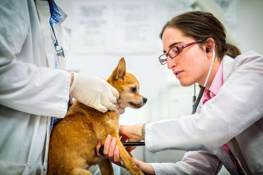
 Your scheduled visit to the Internal Medicine Service at the Cornell University Hospital for Animals begins with a check-in at the reception desk. Following a small amount of paperwork, you will be greeted in the waiting room by one or two students rotating on the Internal Medicine Service and be taken to a private examination room. Once there, the students will collect information relative to the history of your pet’s health and then perform a physical examination. We appreciate your patience and understanding in allowing these future veterinarians to interact with you and your pet.
The students will then consult with the Internal Medicine resident and faculty regarding their findings and assessments. The resident will accompany the student back into your examination room and will examine your pet again and then discuss a plan to reach a diagnosis, including optional next steps, cost and logistics. Although we encourage our residents to take the lead on first stage assessments and interactions, they are always required to consult with a faculty member to develop a plan for further diagnosis and treatment.
Often, you will be asked to leave your pet in our care such that we can begin appropriate testing. Given our schedule and consultations with other specialists regarding your pet's care, you may be asked to return to discuss the findings later in the day. Some animals with more serious conditions will be admitted to the hospital for further monitoring and treatment.
Finn's Fund for Dogs With Liver Abnormalities
Finn’s Fund was created to help animals, and their families with financial limitations, clarify the diagnosis and medical care if they are suspected or confirmed to have hepatic vascular abnormalities. Eligibility will be based on recommendation from a referring DVM through Drs. Sharon Center or John Loftus in the Small Animal Internal Medicine Service at Cornell. Qualifications include financial need, hopeful prognosis for quality of life commensurate with the typical household family companion, treatment compliance and dedication to medical management of the disease.
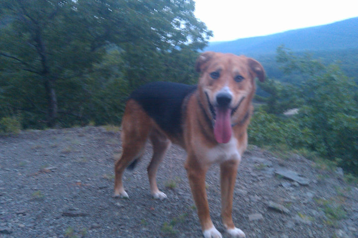
In 2006, Finn was adopted by Christina Fried '94 and became a beloved member of her family. Unfortunately, at a very young age, he started exhibiting signs of serious and chronic illness. Just before his second birthday, through a liver biopsy, Finn was diagnosed with Primary Portal Vein Hypoplasia (PPVH) with portal hypertension. PPVH cannot be cured, but Christina was committed to finding treatment to help hold the disease at bay, manage the symptoms, and ward off complications. The medical management of Finn's health was complicated, time intensive, and costly. Through her tenacious dedication to Finn and finding him the best care available, they were able to share an additional 9 years together.
Christina established Finn's Fund to assist other families whose dogs are facing a vascular liver abnormality. She knows how difficult it can be to get an accurate diagnosis, let alone manage the costs associated with treating the disease. She wants to ensure that families have access to top-notch care, allowing them to make the necessary treatment choices without financial hardship.
"Finn was a constant reminder to myself and those around him of all of the good and wonderful things that life can bring. Despite being abandoned as a puppy to the streets of New York, he chose to see the world around him as full of love, excitement and wonder. Though he had to spend far too much time in the hospital, he always bounded up the stairs with a wagging tail and kisses for everyone. He brought such happiness and joy to all the people he encountered, and I truly believe that he made us become better people. Although his absence leaves a big hole in all of our lives, I feel so lucky to have been able to share his life and so grateful for every day that we were able to extend it through the treatment designed by his doctors and Cornell." – Christina Fried '94
For more information or to be considered for this fund please Contact Us

Related Info
American College of Veterinary Internal Medicine
A non-profit board created to enhance animal and human health by advancing veterinary internal medicine through training, education, and discovery.