

Ophthalmology

 The Ophthalmology Service at the Cornell University Hospital for Animals provides scheduled and emergency care for companion animals with eye and vision problems. Our staff include the board-certified ophthalmologists and residents who collaborate with other veterinarians across the Northeast to provide comprehensive eye care.
Our experienced veterinarians offer the latest diagnostic and treatment techniques using a state-of-the-instrumentation. We offer advanced diagnostics and we utilize advanced surgical techniques. We work collaboratively with other services in the hospital to provide a comprehensive diagnosis and treatment of the full spectrum of veterinary ophthalmic disorders.
Advanced Techniques
- Electroretinography
- Ocular ultrasonography (standard ocular and high resolution anterior segment scans)
- In vivo corneal confocal microscopy
- CT and MRI scans
Surgical Services
- Orbital and adnexal surgery
- Corneal therapeutic and reconstructive procedures, keratotomies, grafts, and transpositions
- Cataract removal by phacoemulsification with intraocular lens implantation
- Glaucoma management
- Diode laser retinopexy and uveal neoplasia and cyst ablation
What to Expect During Your Appointment
 Your scheduled visit to the Ophthalmology Service at the Cornell University Hospital for Animals begins with check in at the reception desk. Following a small amount of paperwork, you will be greeted in the waiting room by one or two students currently in their ophthalmology rotation and brought to a private examination room.
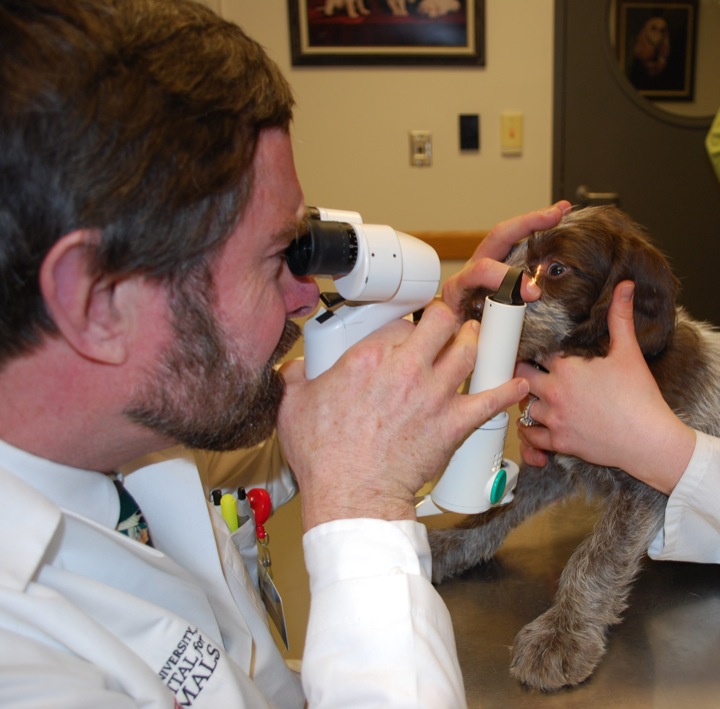
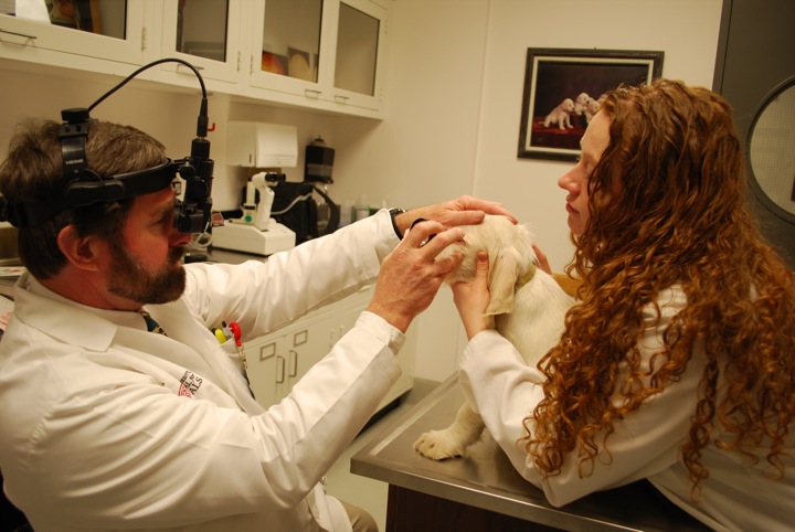
The students will conduct an examination of your pet and ask you questions about your animal's history and current health. In the course of this examination, several tests are usually performed including measurements of tear function and eye pressure. This initial evaluation will take 30-45 minutes. We appreciate your patience and understanding in allowing these future veterinarians to interact with you and your pet.
The student will then administer dilating eye drops, which take 15 to 20 minutes to take effect. During that time, the student will then leave to consult with a resident or faculty member to analyze the results of the routine tests and refine plans to further diagnose and treat your pet's eye conditions.
The student will return to complete the dilated portion of the examination. Then an ophthalmologist will perform a second comprehensive examination and discuss the findings with you. Together, you will develop a plan for further diagnosis and treatment. Most patients that require surgery or advanced tests such as CT or MRI will be admitted to the hospital from their initial appointment.
 OPHTHALMIC EMERGENCIES:
All daytime and after-hour ophthalmic emergencies are admitted and managed directly by the faculty and resident clinicians of the Ophthalmology Service.
Ophthalmology: Medical Conditions
 Common Ophthalmic Conditions
 Common Ophthalmic Conditions
Corneal ulcers
The surface of the eye is commonly injured and usually heals spontaneously without treatment or predictably with supportive treatments. But these injuries may result in ulceration of the cornea, which may become infected by bacteria or become deep enough to threaten loss of the vision and the eye. Diagnosis is made by complete ophthalmic examination. Treatment involves topical antibiotic therapy supported by surgical intervention to stabilize deep ulcers or wounds. With early aggressive treatment, outlook for healing is generally good.
Glaucoma
Glaucoma is increased pressure within the eye leading to permanent vision impairment. It is an inherited condition in many breeds of dogs and some cat breeds. It also occurs secondary to other ocular disorders such as inflammation, tumors, trauma in all species.
The outlook for preserving vision in eyes with inherited glaucoma is guarded, with early diagnosis and treatment yielding the best results. Diagnosis is made by tonometry - a measurement of intraocular pressure. Management is both medical, using topical pressure control drugs made for people, and surgical, to reduce eye pressure and achieve comfort.
Cataracts
Cataracts develop as common inherited defects in dogs and some other species. Most diabetic dogs develop secondary blinding cataracts. There are no proven or approved medical treatments to prevent, retard, or reverse cataract development, but cataract surgery is performed successfully on animals. Dogs are the most common recipients and most get intraocular lens implants like people do, returning durable useful vision.
Cataract Surgery for Dogs
By definition a cataract is any focal or diffuse opacity of the normally transparent lens. Cataracts are commonly caused by inherited defects of the lens, metabolic disorders (most commonly diabetes mellitus), and traumatic injuries. Many, but not all cataracts, progress in one or both eyes to cause vision impairment and blindness. Pets with cataracts can be evaluated for cataract surgery and have the surgery performed at the Cornell University Hospital for Animals by a veterinary ophthalmologist.
Appointments for cataract surgery evaluations are scheduled for Monday and Tuesday mornings. Prior to this appointment, we recommend that your dog have a complete physical examination by your veterinarian and two blood tests (a complete blood count and chemistry panel) and a urinalysis performed within one month before the appointment. The results of these should be brought with you to the appointment. After a complete eye examination is performed on your pet, the procedures involved in cataract surgery will be discussed with you. In most instances, dogs can be admitted from this appointment for surgery that same week. Most dogs are hospitalized for three to four days. Priorto the surgery, two additional tests will be performed: electroretinography (ERG) and an ultrasound examination. The ERG assesses the function of the retina, the light-sensitive layer of the eye; the ultrasound examination looks for retinal detachment. If retinal function is poor by ERG determination or if the retina is detached, surgery may not be performed.
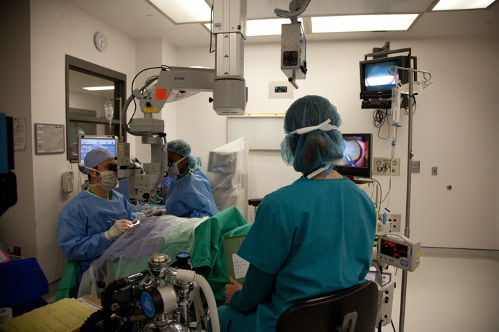
Cataract surgery is performed under general anesthesia, usually on both eyes at the same time. An intraocular lens (IOL) is usually inserted after the cataract has been removed. The success rate of uncomplicated cataract surgery is 85 to 90%. Postoperative concerns include excessive postoperative inflammation, bleeding, glaucoma (increased eye pressure), and retinal detachment. Note that these complications are also common in eyes with blinding cataracts that are not operated on!
Because dogs’ eyes develop more serious inflammation than human eyes after cataract surgery, they must receive treatments (a combination of pills, eye drops, and ointments) several times daily for four to six weeks after this surgery. They also must be rechecked by a veterinary ophthalmologist two or three times during this period. Both postoperative treatments and follow-up are critical to achieve the best results!
The cost of uncomplicated cataract surgery is approximately $3,675, inclusive of the preliminary examination, ERG and ultrasound examinations, hospitalization, initial medications, surgery, anesthesia, and operating room use. The professional fee for the first three postoperative rechecks within 90 days is included in the surgery fee; medication refills are not included.
Note: If dogs are receiving cortisone drugs (e.g., prednisone, dexamethasone) for skin or other conditions, or arthritis drugs (e.g., Deramaxx ®, Rimadyl ®, Zubrin ®, Aspirin), these must be stopped at least 10 days prior to the appointment.
To schedule a consultation for your pet with cataracts with the Ophthalmology Service, please call the Companion Animal Hospital at the College of Veterinary Medicine at Cornell University at (607) 253-3060. New York State Law requires a valid rabies certificate be presented upon arrival to our hospital. Failure to do so may result in your pet not being evaluated.
Uveitis
This inflammation within the eye is commonly associated with ocular and systemic infections, immune-mediated and metabolic disorders, and is a frequent cause of temporary and permanent vision loss. Treatment and prognosis depend upon stage at diagnosis, causes, and control of associated medical conditions. Diagnosis is based upon complete ophthalmic examination and treatments include topical and systemic anti-inflammatory medications.
Retinal degeneration
Retinal degeneration is common in dogs and some other species. It is often an inherited defect but also can result from exposure to certain drugs or chemicals. In dogs, it may occur suddenly for unknown reasons. It is diagnosed by complete ophthalmic examination . Few retinal degenerations are treatable; most eventually result is severe vision impairment and blindness.
Related Info
American College of Veterinary Ophthalmologist
A non-profit organization that certifies veterinarians in ophthalmology and provides information about veterinary eye disorders.