

Border collie bounces back after rare ligament injury
Rook-4-PC-Provided-resize.png
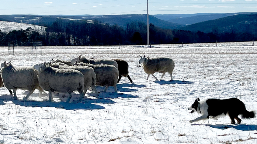
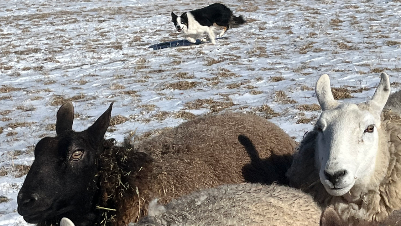
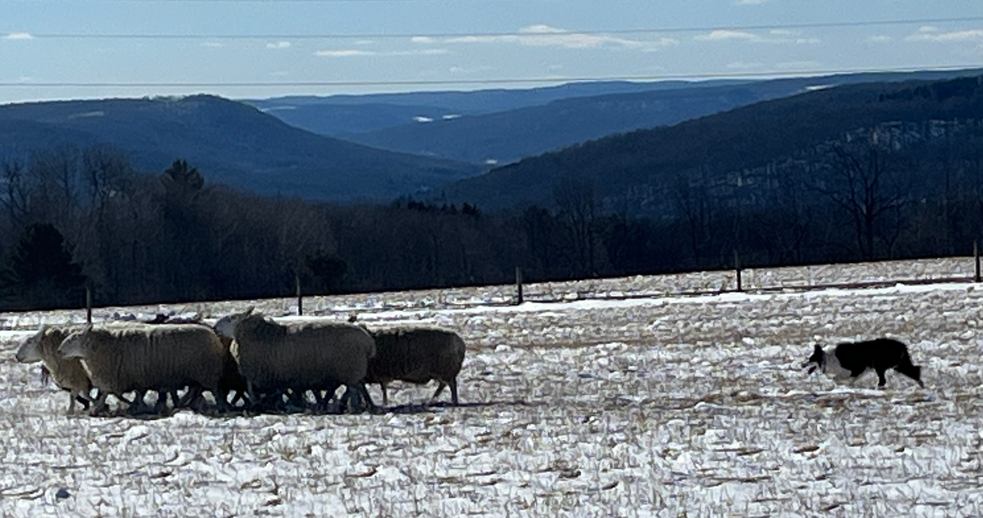
Border collie Rook at work herding sheep. Photo provided.
At the top of the Appalachian plateau outside Rochester, it’s not uncommon to see a flash of black and white fur dashing through the sprawling vista. This is Diane Cox’s home, and that furry meteor is Rook, one of her border collies and a champion herder. One day, however, when Rook’s leg collided with the bottom of a gate hidden by tall grass, it seemed like his promising herding career had ended before it truly began.
“He was going at a dead sprint. I thought this was it, that his competitive career was over,” Cox said.
For a working dog like Rook, not being able to herd would be a major change in his daily life. Cox called the Cornell University Hospital for Animals for an appointment with the Sports Medicine and Rehabilitation Service to explore his treatment options.
Section chief Christopher Frye, D.V.M. ’11, and first-year resident Karolynn Ellis ’18, D.V.M. ’22, led Rook’s care team. They found that Rook had a traumatic caudal cruciate ligament tear — a ligament within the knee known in humans as the PCL. “A tear like this results in knee pain, swelling and a degree of knee instability with the progression of eventual osteoarthritis,” Frye said. The PCL provides knee stability, particularly against rotational forces. It also provides spatial awareness feedback for fine neuromuscular control.
“This is particularly concerning for Rook, a herding dog that requires tight turns at high speeds,” Frye said.
Experiencing the warmth and care extended to herself, Rook and others in the hospital waiting room made a big impression on Cox. She has been part of the competitive herding world for 10 years and has traveled with her dogs across the country to participate in national events with much success. Rook showed promise as a competitor from an early age. “He’s a thinker,” Cox said. “He has so much presence and is cool as a cucumber when he’s working. I could send him out of sight and trust him to make the right call.”
Treatment plan
Usually, the PCL is torn when its companion, the cranial cruciate ligament — in humans, the ACL — ruptures. Rook’s case was particularly uncommon because his ACL remained intact and healthy; only the PCL was injured. In dogs, the ACL is commonly diseased and leads to severe weight bearing instability of the knee as well as rotational instability. In canine medicine, we have developed surgeries such as a TPLO (Tibial Plateau Leveling Osteotomy) to correct that weight bearing instability because biological grafts, like in human medicine, fail. The PCL injury does not result in the dramatic weight bearing instability we see in ACL tears due to the weight bearing axis and bony conformation of dog knees. Because there are no well-established surgical solutions for PCL injuries in dogs, medical management is the mainstay of treatment.
“First, we used ultrasound to verify the cranial cruciate ligament was healthy, and then proceeded with the MRI to diagnose a ruptured PCL,” Frye said.
To treat the injured ligament, Rook’s care team used platelet rich plasma therapy. Under sterile conditions and guided by an ultrasound, platelet rich plasma is injected into the joint, where it reduces inflammation and pain. This was followed by staged rest and months of physiotherapy targeted at strengthening and coordinating the muscles that help stabilize the stifle; particularly against rotational forces. Rook returned to Cornell at several intervals for treatment, with the hope that he could soon return to the sport he loves. “His veterinarians were a delight every time we came to Cornell,” Cox said.
It was important for Rook to keep his legs strong between injections. Fortunately, Cox’s sprawling property was perfect for rebuilding muscles around his knee. This gave his ligament the best chance for healing without relying on surgery.
“Rook’s case was a good example of how multiple imaging techniques, like ultrasound and MRI, can provide a definitive diagnosis without more invasive steps like exploratory surgery,” Frye said. “We are incredibly excited to see Rook doing so well.”
Indeed, within six months, Rook was fully recovered and back to competitive herding.
A healing journey
Cox has a special bond with Rook, whom she describes as the smartest dog she’s ever had. However, it isn’t for this reason alone that his recovery has meant so much to her. Cox was recently diagnosed with an aggressive form of multiple myeloma, a blood cancer, and is passionate about doing what matters to her, like training and competing with Rook.
“So in a way, they helped not just my dog, but me,” Cox said.
Cox and Rook will soon climb into her Airstream and head west for more national competitions, where they’ll meet up with other members of the herding community. “I’m revisiting the most gorgeous landscapes of my childhood — and doing the things that matter while I’m there,” Cox said.
Rook’s entire Cornell team is pleased to have made such a difference. “We enjoyed working so closely with Rook and his mom,” Frye said. “We feel a great bond with our patients and their families, and nothing makes us happier than achieving their goals, and watching Rook get back to sport.”
Written by Melanie Greaver Cordova


